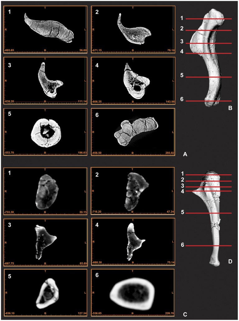
Figure 5. CT scan images of humerus and ulna displaying cross-section morphology.
Humerus cross-sections images 1–6 (A), Computer generated render of humerus with corresponding image cross-section lines 1–6 (B), Ulna cross-sections images 1–6 (C), Computer generated render of ulna with corresponding image cross-section lines 1–6 (D). Scale is included as part of each CT image in mm.

