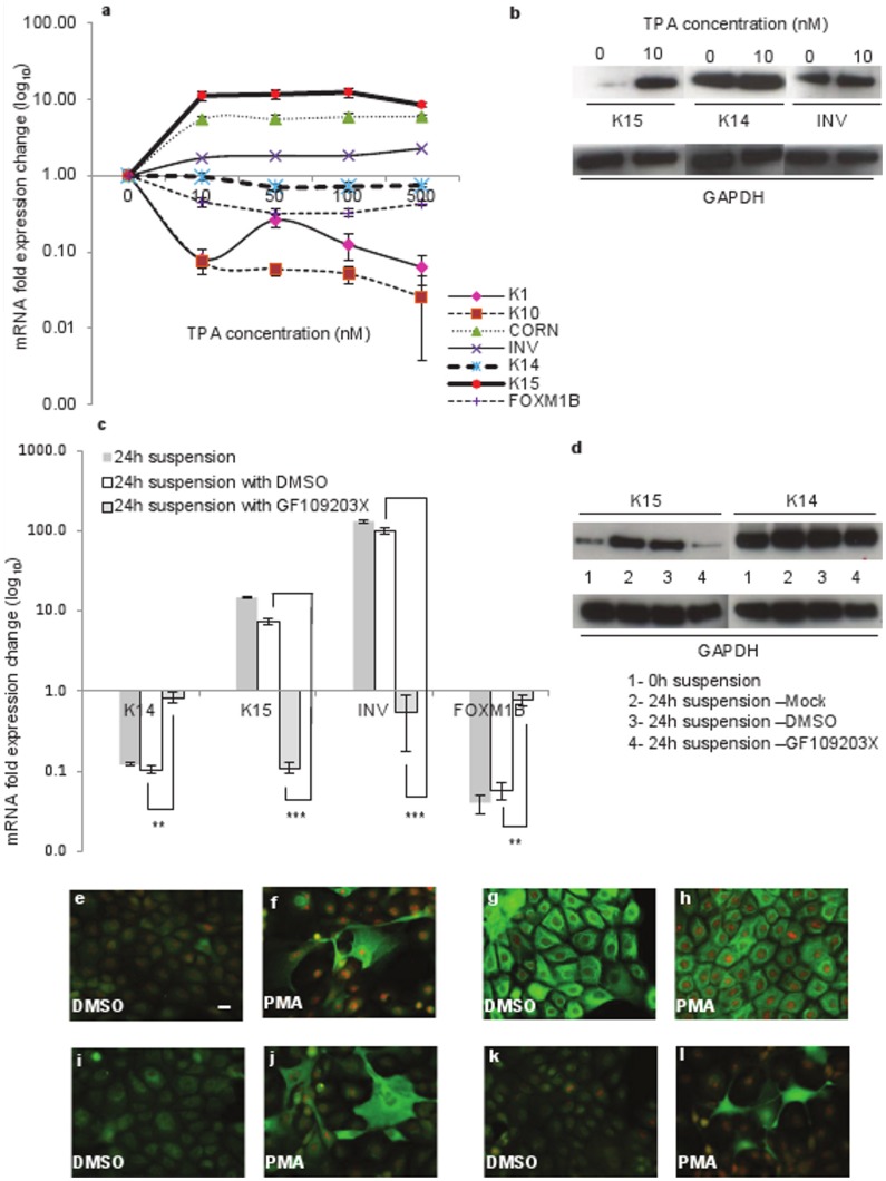
Figure 5. Influence of PKC activator and inhibitor on the expression of K15 in keratinocytes.
(a) N-Terts in RM+ were exposed to different concentrations of PMA (0–500 nM) or DMSO for 24 h and mRNA expression was determined by qPCR. (b) The keratinocytes were exposed overnight to either 0 or 10 nM PMA, and protein levels were determined by western blotting. (c) N-Terts grown in SFM were suspended in 1.3% (w/v) methylcellulose with no additive, 0.02% DMSO, or 5 µM PKC inhibitor GF109203X for 24 h and the cells were used to determine mRNA expression. Freshly trypsinised single cell suspension of N-Terts was used as 0 h control cells. (d) N-Tert keratinocytes were suspended as in (c) with GF109203X and protein expression at 0 h or 24 h was determined by western blotting. Each bar represents the mean±SEM where n = 3. (P<0.01, very significant, **; P<0.001, extremely significant, ***). For immunostaining N-Terts growing on glass coverslips in RM+ were treated with 10 nM PMA (f, h, j, l) or DMSO control (e, g, i, k) for 24 h. The cells were fixed in 3.8% formaldehyde and immunostained with antibodies against K15 (e, f), K14 (g, h), involucrin (i, j) and cornifin (k, l) (magnification bar = 20 µm).

