
Fig. 7.
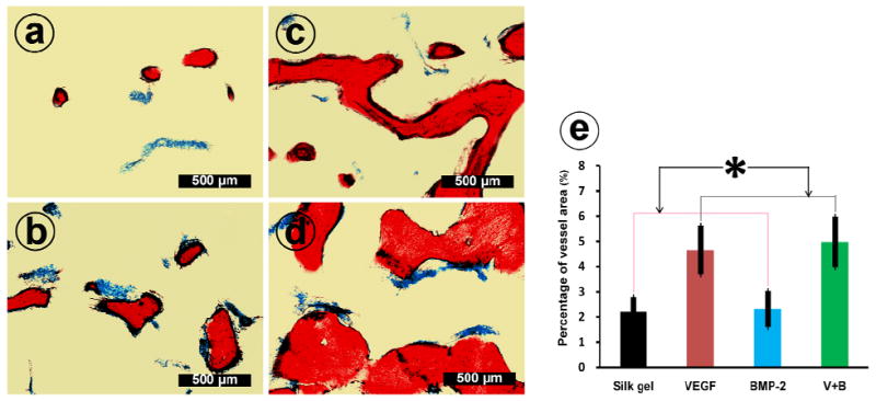
The undecalcified sections of group Silk gel (a), group VEGF (b), group BMP-2 (c) and group V+B (d) at 12 weeks post-operation were further stained with Von Gieson’s picro fuchsin. On those nondecalcified sections, red areas represent newly formed bone and blue areas belong to blue colored Microfil which indicate newly formed blood vessels. (e) This graph shows the percentage of newly formed blood vessel area in the four groups (* indicates significant differences, p < 0.05).