
Fig. 9.
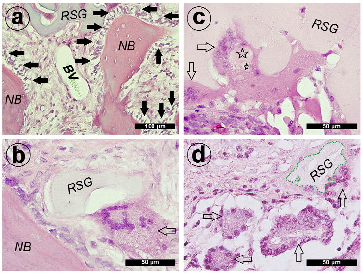
Hematoxylin and eosin staining of decalcified sections of group V+B. (a) New bone (NB) and blood vessel (BV) formation around remnants of the silk gel (RSG). A line of active osteogenic cells was observed around the newly formed bone (solid arrows show active osteogenic cells). At 4 weeks post-operation, (b) multinucleated giant cells quickly invaded into the silk gel and were observed around remnants of the silk gel (blank arrow shows multinucleated giant cell). (c) Silk gel fragments engulfed by the giant cells were also detected (☆ = small silk gel fragments). At 12 weeks post-operation, more giant cells invaded into the silk gel (the boundary of remnants of the silk gel marked with green dash line; blank arrow shows multinucleated giant cells).