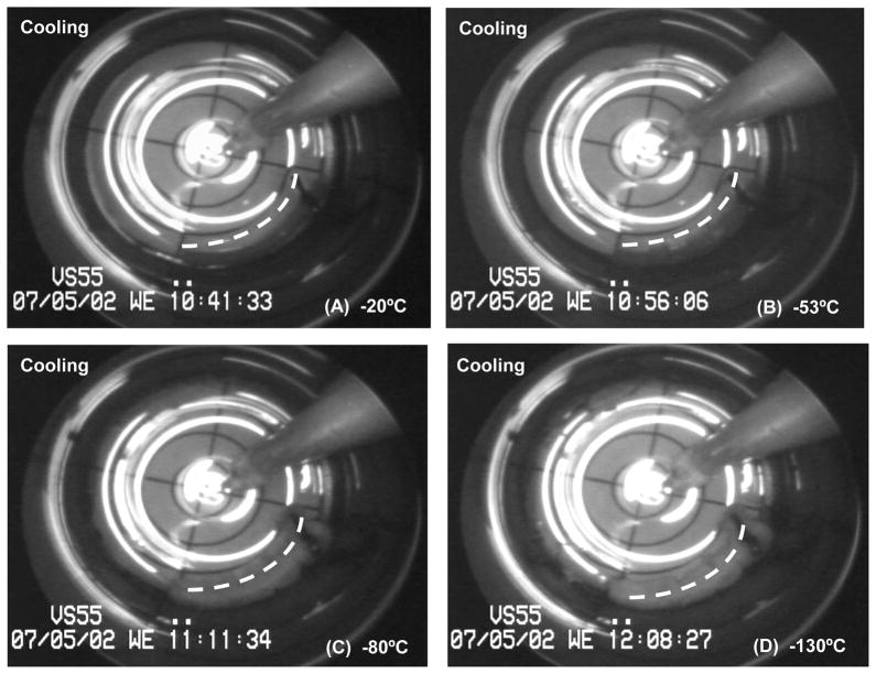
Figure 5.
Cryomacrographs of so-called “negative control” vitrification. The vessel centerline is represented by a dashed white line. Viability testing detected no mechanical function and extremely low metabolic activity post slow cooling/slow rewarming cryopreservation. Fractures were seen (Panel D) on the tissue opposite side of the vial and on the lower left side of the vial, near the suture end of the vessel segment.

