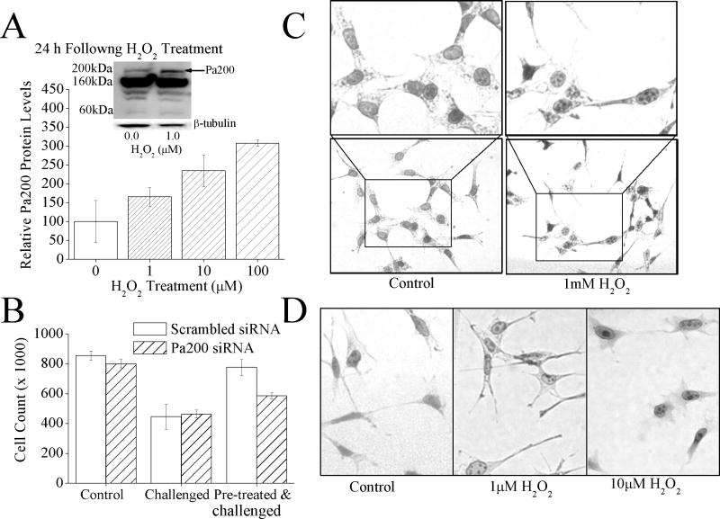
Fig. 3.
(A) H2O2 exposure causes an increase in Pa200 protein levels. MEF cells were pretreated with 0, 1, 10, or 100μM H2O2, 24 h later samples were harvested and run on Western blots which were then screened with antibodies against either Pa200 or the loading control β-tubulin. Sample band intensity was measured and adjusted based on β-tubulin band intensity. Values are means ± SE where n = 3. The inset shows a representative blot. (B) By blocking the adaptive increase in Pa200, the H2O2 induced increase in oxidative stress tolerance is blunted. MEF cells treated with either Pa200 or (scrambled) siRNA for 24 h to block induction. Samples were then transiently adapted to oxidative stress by pre-treatment with 1μM of H2O2, for 1 h. Following a 24 h adaptation period, both adapted and non-adapted cells were challenged by incubation with a high dose of 100μM H2O2. Cell counts were then taken using a cell counter 24 h later. Results are means ± S.E. where n = 3. (C) Exposure to a toxic level of H2O2 causes the formation of Pa200 foci in the nucleus. MEF cells were exposed to 1.0mM H2O2 for 1hr. Immunocytochemistry was then performed and cells were stained with anti-Pa200. (D) Exposure to low, adaptive doses of H2O2 also results in the formation of nuclear Pa200 foci. Conditions were identical to those of panel C., except that 0, 1.0μM, or 10.0μM H2O2 exposures were used.

