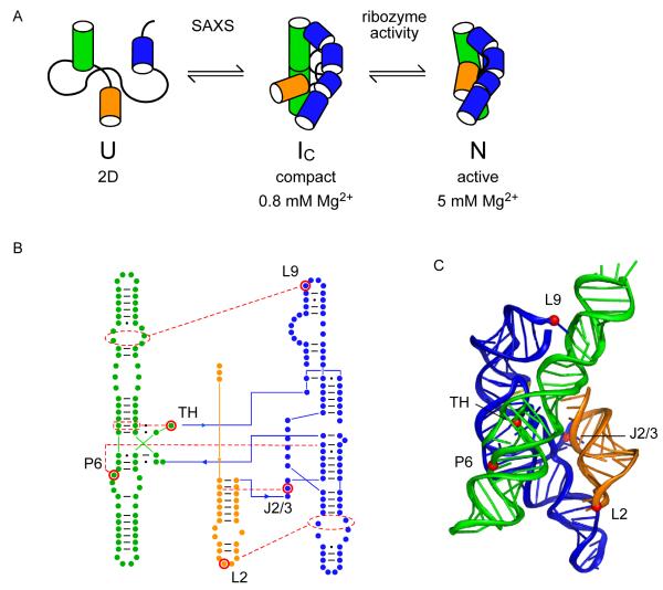
Figure 1. Cooperative folding of the Azoarcus group I ribozyme.
(A) Compact, native-like intermediates (IC) form in low Mg2+ (Perez-Salas et al., 2004; Rangan et al., 2003) and are detected by SAXS or native PAGE. Formation of the native structure (N) is reported by ribozyme activity and the solvent accessibility of the RNA backbone. See also Figure S1. (B,C) Tertiary interaction motifs indicated by red dots were perturbed by single base substitutions: loop L2, A25U; joining region J2/3, A39U; paired region P6, A97U; triple helix TH, G125A; loop L9, A190U (see Table S1). J8/7, cyan ribbon. Cooperative interactions indicated by red (IC) or blue (N) arrowheads (positive, pointed; negative, flat). Thickness indicates relative strength. (C) Ribbon drawn with PyMOL; 1u6b (Adams et al., 2004b).

