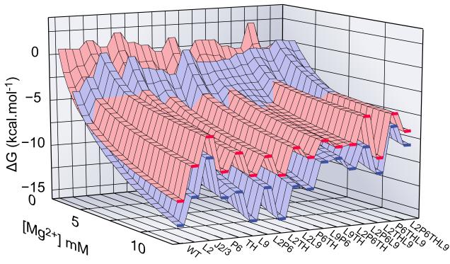
Figure 4. Free energy landscape for folding.
Energetic perturbations in the Azoarcus ribozyme due to mutations in IC (red) and N (blue) are shown versus Mg2+ concentration (back to front). Mutations that destabilize N more than IC, e.g. J2/3 and most of triplets, shift the crossover of surfaces to higher Mg2+ compared to WT. Red and blue surfaces are similar because IC is similar in structure to N. Single mutations located in helical junctions (J2/3 and TH) are particularly destabilizing. See also Figure S4.

