
Fig. 1.
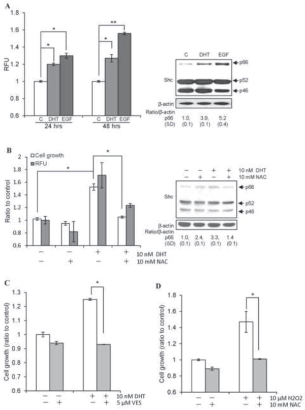
Androgens upregulate ROS, p66Shc protein and PCa cell proliferation. (A) DHT and EGF increased ROS production in LNCaP C-33 cells. Cells were plated at a density of 5×10³ cells/cm2 in duplicates for 3 days in regular RPMI medium. Cells were steroid starved for 48 h in steroid-reduced (SR) medium, and then treated with 10 nM DHT or 10 ng/ml EGF for the indicated time periods. Cells were then incubated with 20 μM dichlorofluorescein diacetate (DCF-DA) for 15 min and ROS levels were measured by flow cytometry. Median fluorescence was calculated and normalized to the control values and expressed as Relative fluorescence units (RFU). (left panel, *p<0.05; **p<0.01). Total cell lysate proteins from 48 h DHT or EGF treatments were analyzed for Shc protein. β-actin protein level was used as a loading control (right panel). (B) LNCaP C-33 cells were seeded and followed by steroid starvation as described above. Cells were then treated with 10 nM DHT in the presence or absence of 10 mM NAC for 48 h. The cell growth was determined by cell counting and ROS levels were measured as described above. The data shown is an average of three sets of independent experiments in duplicates (left panel, n=2×3, *p<0.01). Total cell lysate protein was analyzed for Shc protein. β-actin protein level was used as a loading control (right panel). (C) MDA PCa2b cells were plated at a density of 1×10⁴ cells/cm2 in duplicates for 3 days in regular medium. Cells were steroid starved for 48 h in SR medium and then treated with 10 nM DHT in the presence or absence of 5 μM VES for 48 h. The cell growth was determined by cell counting. The data shown is one set of representative results from two sets of independent experiments (*p<0.05). (D) LNCaP C-33 cells were seeded and followed by steroid starvation as described above. Cells were then treated with 10 μM H2O2 in the presence or absence of 10 mM NAC for 48 h. The cell growth was determined by cell counting (*p<0.05).