
Fig. 2.
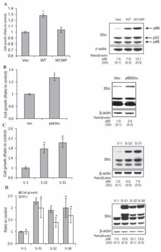
Effect of elevated p66Shc protein expression on PCa cell proliferation. (A) LNCaP C-33 cells were plated in duplicates for 48 h and then transfected with WT p66Shc cDNA (WT). Control cells were transfected with empty vector (Vec) or the redox-defective mutant W134F. After transfection, cells were fed with regular medium for overnight, then steroid-starved for 48 h, and cell growth was analyzed by cell counting (n=2×3, *p<0.01). The data shown is one set of representative results. Immunoblot analysis was performed for p66Shc level in respective cells (right panel). β-actin protein level was used as a loading control. (B) MDA PCa2b cells were transfected with WT p66Shc cDNA (WT) or empty vector (Vec). Cells were maintained in regular medium for overnight, steroid-starved for 48 h, and cell growth was analyzed by cell counting (n=2×3, *p<0.01). Immunoblot analysis was performed for p66Shc level in respective MDA PCa2b cells (right panel). β-actin protein level was used as a loading control. (C) LNCaP C-33 stable subclones of WT p66Shc cDNA (S-31 and S-32) and vector-alone (V-1) control cells were plated at a density of 8×10³ cells/cm2 in regular medium for 3 days, replenished with fresh regular medium, and cell growth was analyzed by cell counting after 2 days (n=2×3, *p<0.01). The total cell lysates were analyzed for p66Shc protein levels by immunoblotting (right panel). β-actin protein level was used as a loading control. (D) As described above in (C), WT p66Shc cDNA stable subclones (S-31, S-32 and S-36) and control vector-alone (V-1) cells were plated in regular medium for 3 days. Cells were steroid-starved for 48 h, replenished with fresh SR medium for 3 days. Cells were then incubated with 20 μM DCF-DA for 30 min, and trypsinized for counting cell number or measuring ROS levels by flow cytometry (n=2×3; *p<0.01). Total cell lysates were analyzed for p66Shc protein levels by immunoblotting (right panel). β-actin protein level was used as a loading control.