
Fig. 4.
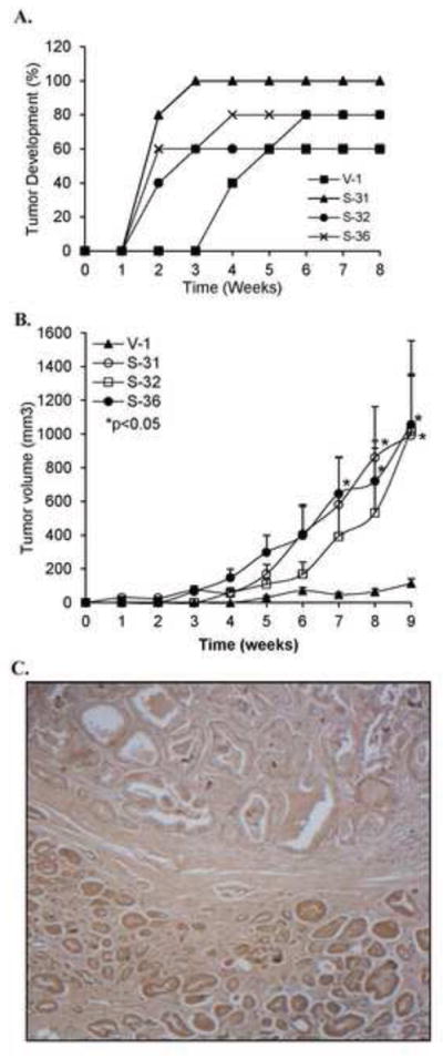
Effect of elevated p66Shc protein expression on PCa tumorigenicity. (A) p66Shc cDNA transfected stable subclone cells (S-31, S-32 and S-36) and control V-1 cells with 1×10⁶ cells each in 0.1 ml medium plus 0.1 ml Matrigel were injected into 5 male nude mice per group. (B) 2×10⁶ cells each in 0.1 ml medium plus 0.1 ml Matrigel were injected into 5 female nude mice per group. The tumor growth was monitored by a caliper weekly. Tumor volume was shown as Mean ± S.E. (S-31: 8 & 9 weeks; S-36: 7, 8 & 9 weeks vs. V-1, *p<0.05) (C) Immunohistochemical staining for p66Shc protein in human prostate tissue archival specimen using anti-p66Shc antibody. Note the prostatic adenocarcinoma (bottom) with higher levels of p66Shc protein as compared to benign prostatic acini (top). Original magnification x200.