
Fig. 5.
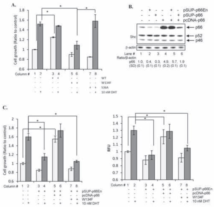
p66Shc via ROS mediates DHT-stimulated cell proliferation. (A) LNCaP C-33 cells were plated at a density of 1×10⁴ cells/cm2 in duplicates. Cells were transfected with either WT, W134F mutant or S36A mutant cDNA of p66Shc. Control cells were transfected with pcDNA 3.1 alone. Cells were then steroid-starved for 48 h and treated with either ethanol or 10 nM DHT. Cell numbers were analyzed after 48 h of treatment. Data shown is the representative of two sets of independent experiments (*p<0.05). (B) Immunoblot analyses were performed for p66Shc protein levels in LNCaP C-33 cells transiently transfected with either shRNA against endogenous p66Shc (pSUP-p66En), p66Shc-specific shRNA (pSUP-p66) or p66Shc cDNA (pcDNA-p66). (C) As described as (A), LNCaP C-33 cells in duplicates were co-transfected with shRNA against endogenous p66Shc (pSUP-p66En) and WT or W134F mutant of p66Shc cDNA. Control cells were transfected with pSUP or pSUP-p66En alone. All cells were further co-transfected with pECFP cDNA to normalize the transfection efficiency. Cells were then steroid-starved for 48 h and subsequently treated with ethanol or 10 nM DHT. Cell proliferation was performed by counting the cells. Results were normalized to the control pSUP vector-transfected cells treated with ethanol. (left panel). ROS was measured after 48 h of DHT treatment using DCF-DA assay. Relative fluorescence units (RFU) were normalized to pECFP fluorescence to nullify the differences in the transfection efficiency (right panel). Data shown is the Mean ± S.E. of one set of representative results from two sets of independent experiments (*p<0.05).