
Fig. 6.
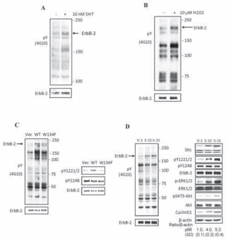
DHT, H2O2 and p66Shc activate ErbB-2 tyrosine phosphorylation in AS PCa cells. (A) LNCaP C-33 cells of 8×10³ cells/cm2 were seeded in regular medium for 48 h. After steroid starved for 48 h, cells were treated with 10 nM DHT and harvested after 24 h. Immunoblotting analyses were performed on the total lysate with anti-Tyr(P) antibody (4G10). After stripping, the membrane was re-hybridized with anti-ErbB-2 protein Ab (lower panel). Arrow marks the position of ErbB-2 protein. (B) LNCaP cells were seeded in regular culture medium and then maintained in a SR condition for 48 h. Cells were fed with fresh SR medium and exposed to 10 μM H2O2 for 16 h. Total cell lysate proteins were analyzed for the total tyrosine phosphorylation profile. The membrane was stripped and then re-hybridized for anti-ErbB-2 Ab (lower panel). Arrow points out ErbB-2 position. (C) LNCaP C-33 cells were transiently transfected with WT p66Shc or its W134F redox-defective mutant cDNA. Control cells were transfected with vector alone (Vec). Cells were steroid starved for 48 h and then harvested. The total tyrosine phosphorylation profile (left panel) and the ErbB-2 site-specific phosphorylation (right panel) were analyzed by anti-p-Tyr Ab (4G10) or Ab to the site-specific ErbB-2 phosphorylation. Arrow (left panel) indicates the position of ErbB-2. The ErbB-2 protein was analyzed by immunoblotting after stripping the membrane (lower panel). (D) p66Shc cDNA-transfected LNCaP C-33 stable subclone cells (S-32 and S-31) and vector-alone transfected control cells (V-1) were plated at a density of 8×10³ cells/cm2 in regular medium for 48 h. Cells were harvested and the total cell lysate proteins were analyzed for (left panel) the total tyrosine phosphorylation profile, and (right panel) Shc, cyclin D1 protein, the site-specific phosphorylation levels of ErbB-2, ERK and Akt proteins. Arrow (left panel) marks the position of ErbB-2 protein.