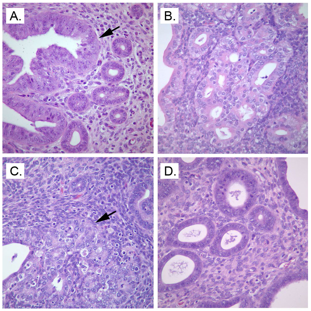
Figure 2.
Mouse endometrium showing (A) endometrium with glandular hyperplasia and atypia in Pten+/− mouse fed the control diet, (B) endometrium with glandular hyperplasia in Pten+/− mouse fed VD3 supplemented diet, (C) endometrial adenocarcinoma in Pten+/− mouse fed the OID, and (D) normal glandular morphology, as in wildtype mice, in Pten+/− mouse fed the OID supplemented with VD3. Endometria were obtained from 28-week-old mice, and sections were stained with hematoxylin and eosin, with 40×objective magnification.

