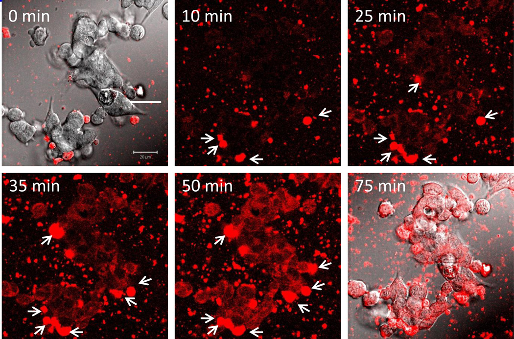
Figure 5. Dynamics and localization of nanozyme transported in exosomes from murine macrophages to CATH.a neurons.
Confocal images of CATH.a neurons incubated with nanozyme (red)-containing exosomes released from preloaded murine macrophages. The first and last time fluorescence images are shown merged with differential interference contrast (DIC) microscopy. Exosomes in the media with encapsulated nanozyme were adsorbed on the surface of the recipient cells (shown by arrowheads) and fused with the membranes, releasing nanozyme into the cytoplasm of neurons that resulted in diffuse fluorescent staining. Bar = 20 µm.

