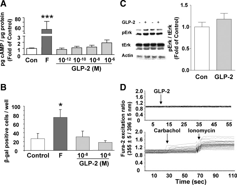
Fig. 4.
GLP-2 does not stimulate cAMP, Erk1/2 phosphorylation, or intracellular calcium in murine ISEMF cells. A, ISEMF cells were treated with media alone [control (Con)], forskolin (F) (100 μm) or h(Gly2)GLP-2 (10−12 to 10−6 m) for 20 min in the presence of IBMX (100 μm), and cell lysates were collected for determination of cAMP by RIA. ***, P < 0.001 vs. control (n = 12–16; L1–L5, at P8, P9, and P11). B, ISEMF cells from CRE-LacZ transgenic animals were treated with media alone (control), forskolin plus IBMX (10 μm each), or h(Gly2)GLP-2 (10−8 and 10−6 m with 10 μm IBMX) for 3 h and then stained for β-galactosidase expression. *, P < 0.05 vs. control (n = 6; L6–L8, at P7 and P9). C, ISEMF cells were treated with 10−8 m GLP-2 for 30 min, and cell lysates were analyzed by Western blotting for the levels of pErk1/2 (pErk) and tErk1/2 (tErk); actin was used as the loading control. A representative blot is shown for two different experiments; graph shows results from densitometry analysis. *, P < 0.05 (n = 6; L2–L6, at P10–P15). D, ISEMF cells were loaded with fura 2 and then treated with 10−10 to 10−6 m h(Gly2)GLP-2. Positive controls included carbachol (100 μm; muscarinic agonist) and ionomycin (1 μm; Ca2+ ionophore). Fluorescent ratio values (355 ± 5/396 ± 5 nm) = (bound Ca2+/free Ca2+) were determined for each cell (n = 98–114 cells; L1, at P5). Arrows indicate addition of treatment; representative experiments are shown.

