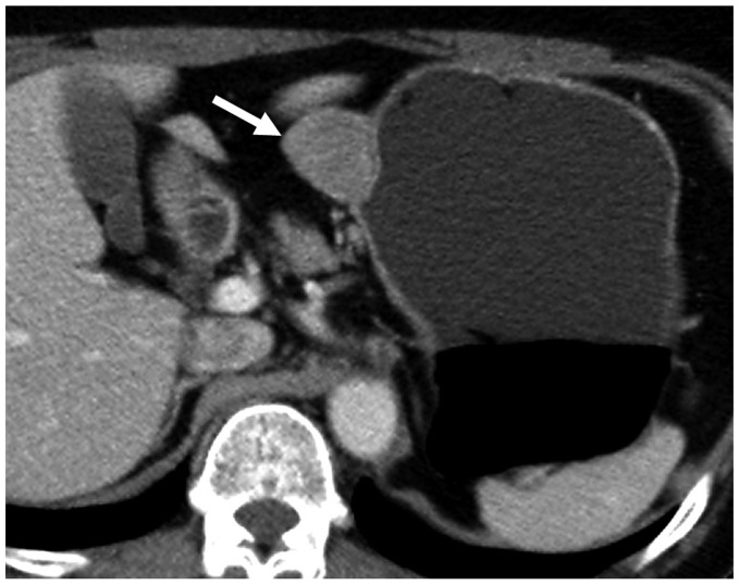
Fig. 2.
Gastric schwannomas in gastric midbody of 62-year-old woman. Transverse CT scan shows well-defined round mass with homogeneously moderate enhancement (arrow) in gastric midbody. Tumor reveals exophytic growth pattern. There is no intralesional low attenuation area, calcification, or surface dimpling.

