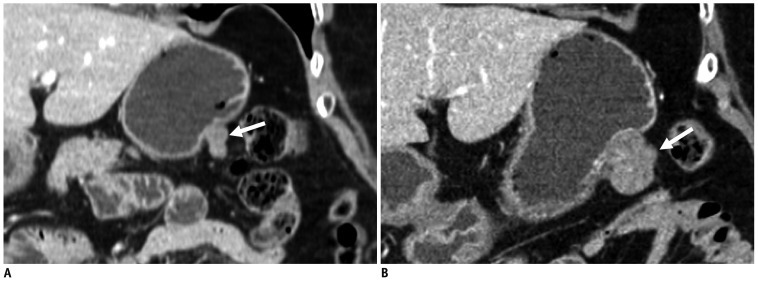
Fig. 7.
Gastrointestinal stromal tumors in gastric upper body of 81-year-old woman.
A. Coronal CT scan shows well-defined lobulated mass with heterogeneous enhancement (arrow) in gastric upper body. Tumor volume is 1610 mm3. B. Coronal follow-up CT scan obtained 12 months after initial CT scan shows 12035 mm3 tumor volume of. This tumor (arrow) has 127-day tumor volume doubling time.

