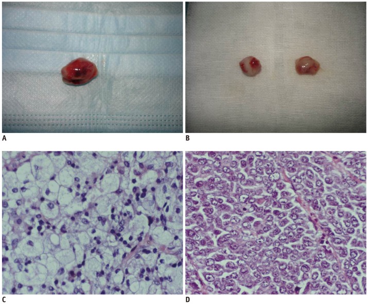
Fig. 1.
Manifestations of gross specimen and histopathological examination of axillary inflammatory and metastatic lymph nodes.
Gross specimen of axillary inflammatory lymph node show that they have smoothly pinkish-grey surfaces and abundant vessels (A), while axillary metastatic lymph nodes appear to have shaggy pale-grey surface with fish like texture (B). Histopathological examination (H & E, × 200) shows inflammatory cell infiltration and cells arrangement sparsely with intercellular substance edema (C), while tumor cells deposit within metastatic lymph node, and enlarged nuclei, hyperchromatism, high nuclear-to-cytoplasmic ratio, and pathological mitotic figure are observed (D).

