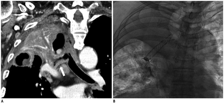
Fig. 1.
77-year-old male (patient 1) with non-small cell lung cancer.
A. Multi-planar reformatted oblique coronal CT image shows bronchial wall thickening with narrowing in right main bronchus and bronchus intermedius (not shown) and total atelectasis of right upper lobe due to obstruction of right upper lobar bronchus. B. Fluoroscopic image shows bronchial stent and aerated right basal lung.

