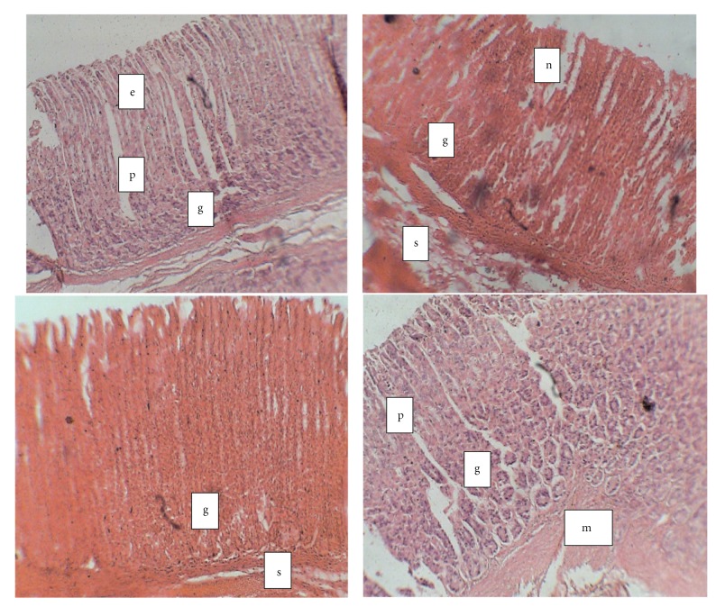
Figure 3.
Photomicrograph of gastric tissue from diabetic and control rats treated with vitamin C (H&E; × 100). (a) Normal control; (b) diabetic control; (c) diabetic + vitamin C; (d) Control + vitamin C. e: epithelium; p: gastric pit; g: gastric gland; s: submucosa; mm: muscularis mucosa; n: necrosis. (a) Stomach from normal control showing: Mucosa is lined by simple columnar epithelium, the gastric pits, underlying gastric glands. The muscularis mucosa and submucosa contain loose areola tissue. (b) Stomach of diabeticcontrol rats showing that mucosa containing tall glandular tissue, the luminal part of which shows some foci of necrosis. (c) Stomach of DM + Vit C showing normal mucosa with tall glandular disposition, no areas of mucosal necrosis. (d) Photomicrograph of the stomach from vitamin C-treated rat showing normal mucosa lined by simple columnar epithelium (e) and numerous gastric glands underneath.

