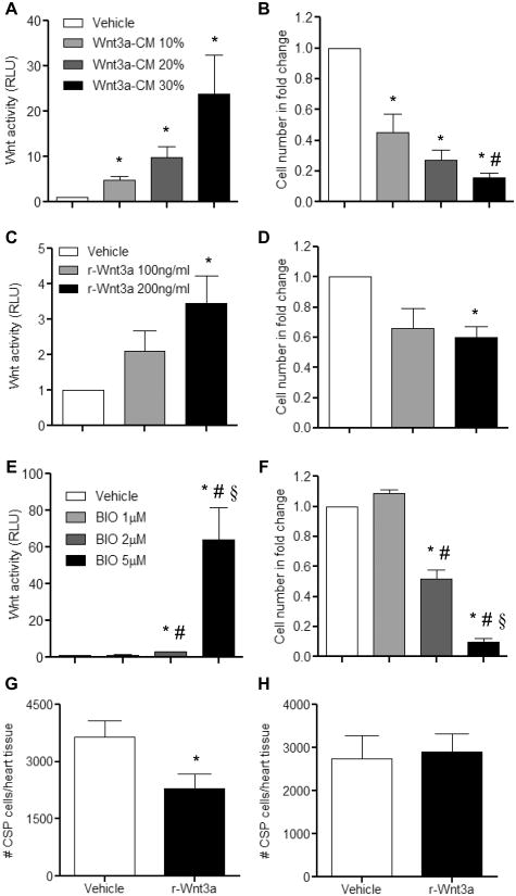
Figure 1. Canonical Wnt signaling pathway mediates anti-growth effects on CSP cells.
Treatment of CSP cells with increasing concentrations of (A) Wnt3a-CM (n=3), (C) r-Wnt3a (n=3), and (E) BIO (n=6) activated the canonical Wnt signaling pathway, as measured by the TCF-luciferase reporter assay. Corresponding proliferation assays using the same dosages of (B) Wnt3a-CM (n=4), (D) r-Wnt3a (n=4) and (F) BIO (n=6) revealed decreased growth capacity of CSP cells. TCF-luciferase activity is measured in relative light units (RLU). Analysis of CSP cell number in (G) the Vehicle or r-Wnt3a injected areas (left ventricular free wall) (Vehicle, n=14; r-Wnt3a, n=16) and in (H) the remote areas from the injection sites (atria, septum, right ventricle) (Vehicle, n=12; r-Wnt3a, n=13). Data are mean ± s.e.m. * P≤0.05 all samples to respective Vehicle, # P≤0.05 Wnt 10% to Wnt 30%, BIO 1μM to BIO 2μM and BIO 5μM, § P≤0.05 BIO 2μM to BIO 5μM. Cell number in fold change presented in B, D, and F is normalized to respective Vehicle group.

