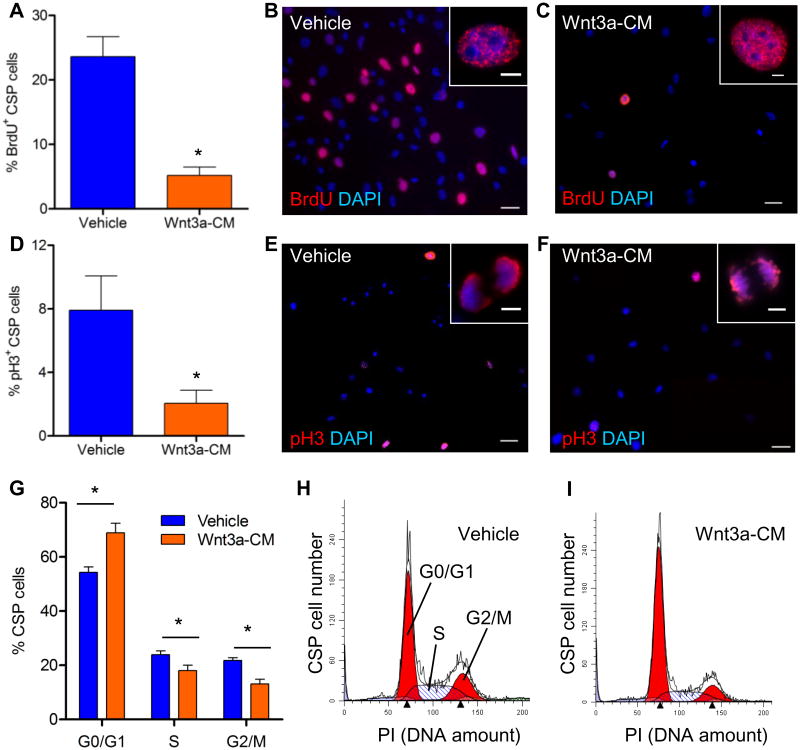
Figure 2. Activation of the canonical Wnt signaling pathway in CSP cells blocks cell cycle progression.
Immunocytochemical analysis of (A) BrdU incorporation (n=5) and (D) expression of p-H3 (n=4) in CSP cells, following treatment with Wnt3a-conditioned-medium (Wnt3a-CM) or Vehicle. Representative images of (B, C) BrdU+ and (E, F) p-H3+ CSP cells, in low (scale bars, 50μm) and high magnification (inset, scale bars, 5μm). (G) Flow cytometric analysis of CSP cells stained with propidium iodide (PI) (n=4). Representative examples of PI analysis following treatment with (H) Vehicle or (I) Wnt3a-CM. Data are mean ± s.e.m. * P≤0.05.

