
Figure 1.
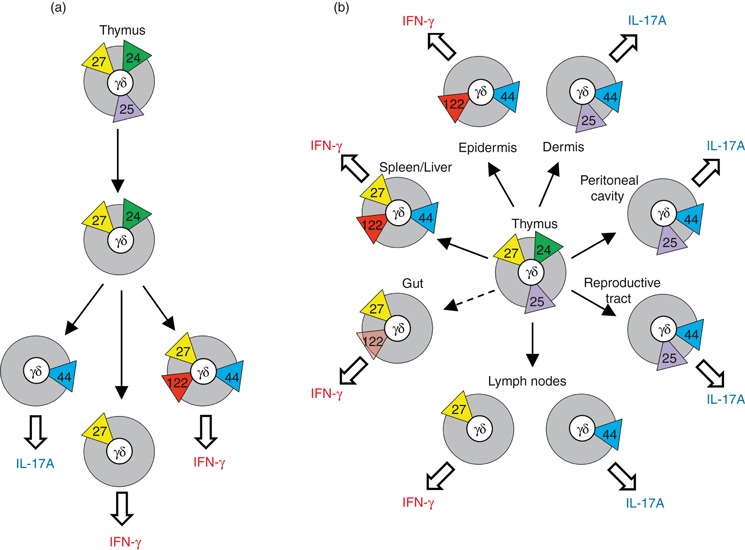
Mouse γδ cell subsets in the thymus and periphery. (a) Thymic γδ subsets described by surface expression of; yellow triangles – CD27; green triangles – CD24; blue triangles CD44; purple triangles – CD25; and red triangles – CD122. Proposed developmental relationships between subsets are indicated by arrows, and potential for cytokine secretion is shown. (b) Peripheral γδ subsets described by tissue location, potential for cytokine secretion and surface markers, as described for (a). Gut γδ cells (i.e. γδ intraepithelial lymphocytes) express only low levels of CD122. IL-17A, interleukin-17A; IFN-γ, interferon-γ.