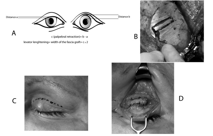
Fig. 3.
Paul Tessier's technique for lengthening the levator muscle of the upper eyelid. This technique is simple, reliable and easily reproduced, and involves a mathematical formula, which can be applied to all patients, to calculate lengthening. The aponeurotic graft is rectangular, and its width is double the palpebral retraction (c) observed before the operation (a). The aponeurotic graft is removed via a coronal (V1) or a temporal (V2) incision to expose the temporal aponeurosis (b). The palpebral incision is made in the superior palpebral fold (c). The upper edge of the tarsal plate is identified and dissected to separate the aponeurosis from the eyelid levator. The aponeurosis is sectioned with a cold scalpel and removed from the muscle. The graft is positioned between the superior edge of the tarsal plate and the inferior edge of the levator and sutured (d).

