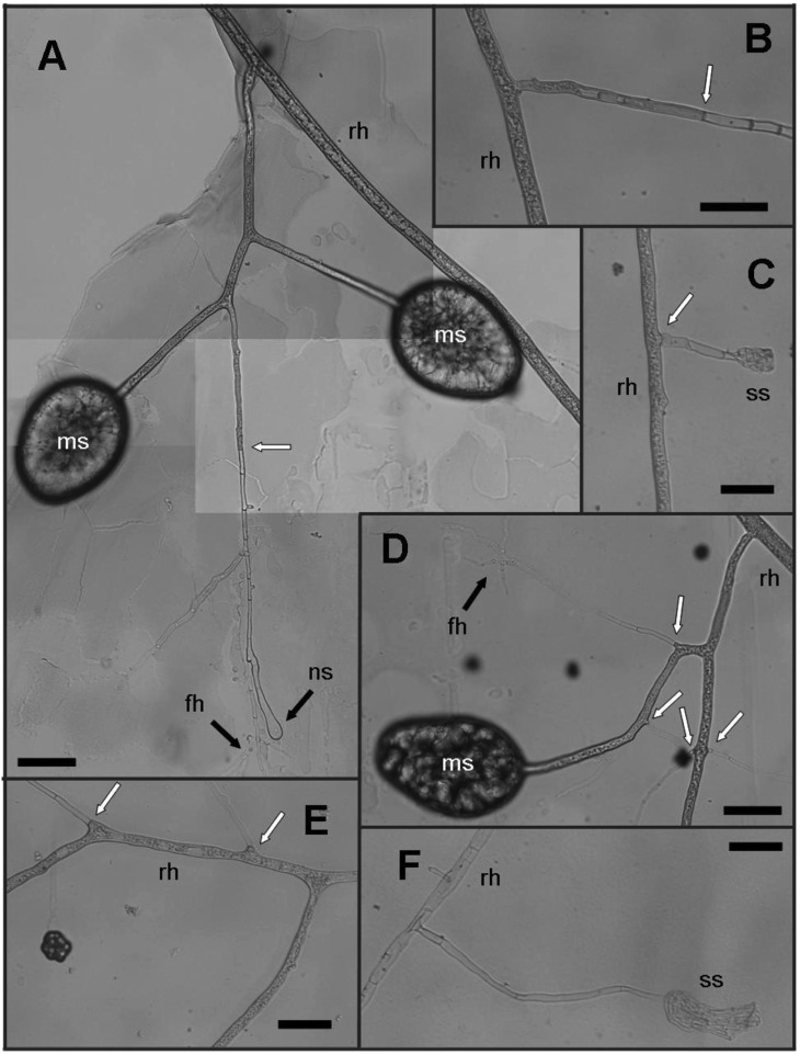
Fig. 3.
Observations of the extra-radical hyphae and spore formation with hematoxylin stain. A, D, Developing spores; B, C, E, F, Different stages of the extra-radical hyphae. Spores (ms) were associated with finely branching hyphae (fh). Cytoplasmic area was distinguished from the non-cytoplasmic area by transparency. A new swollen hyphal apex (ns) formed at a new branch. B, Cytoplasmic contents seemed to migrate from the runner hyphae (rh) into the septate hyphae by removing the septa; C, E, Some of lateral hyphal branches remained septate; C, F, Shrunk-looking structures (ss) were found with the septate hyphae. White arrows indicate the positions where cytoplasm was limited by the septum. Background blots appeared due to the Helly's fluid fixation process (scale bars = 50 µm).

