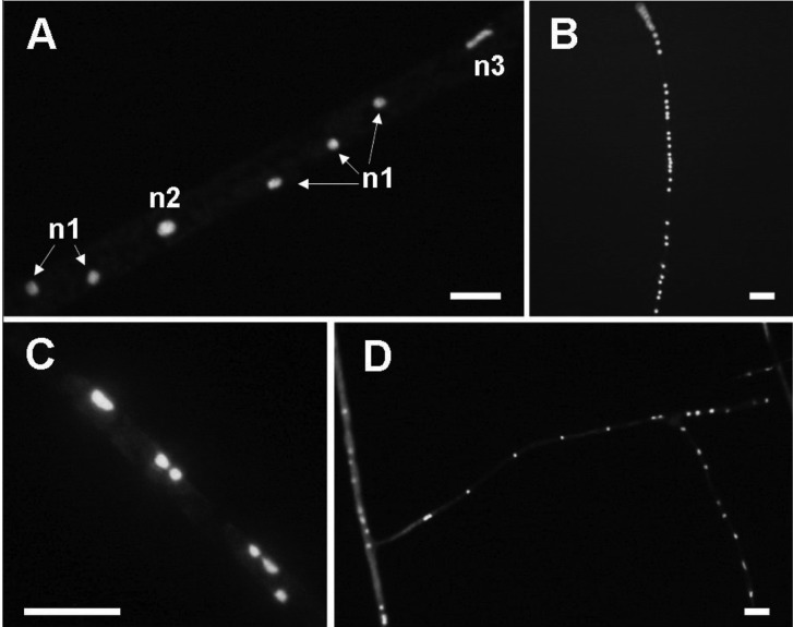
Fig. 5.
Nuclei within a Glomus intraradices mycelium. A, Magnified image of the centre of (E) in Fig. 4. n1, nuclei of usual size; n2, nuclei about twice the normal size; n3, stretched form of nuclei; B, Nuclei existing densely in a row within a thin mycelium; C, Nuclei existing in pairs within runner hypha; D, Nuclei distributed loosely but in regular intervals within a thin hypha (scale bars = 10 µm).

