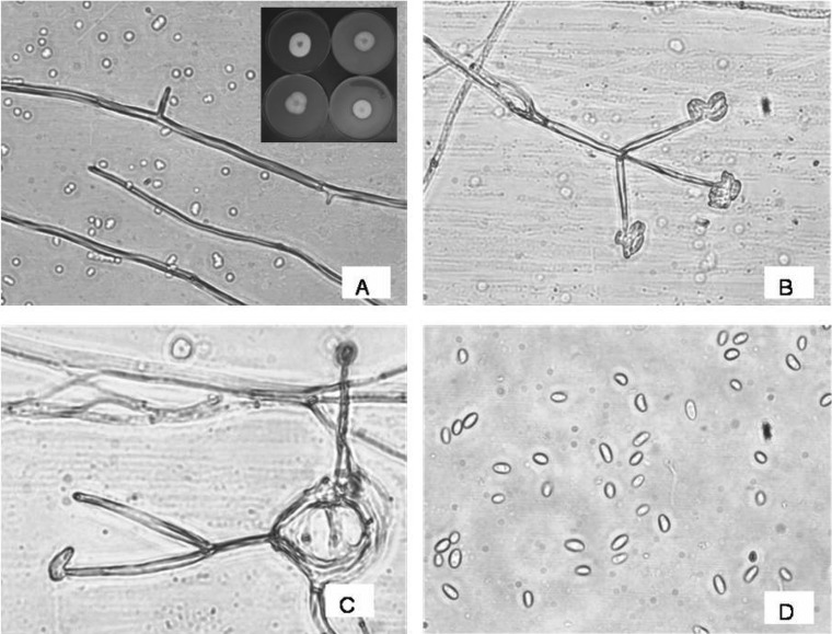
Fig. 1.
Microscopic observations of morphology of fungus AAT-TS-41. A, Mycelium; B, Conidiophores bearing conidia; C, Arrangement of conidia on phialide tip; and D, Conidia. The inset in A showing the picture of Gliocladium sp. growing on potato dextrose agar, oat meal agar, corn meal agar, and V-8 juice (clockwise).

