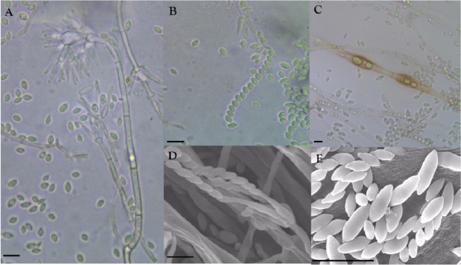
Fig. 2.
Morphological features of the isolate DUCC400. Conidia and conidiophore structures (A), conidia imbricate chain (B), and chlamydospores (C) observed using a phase contrast light microscope. Structures of conidiophore (D) and conidia (E) observed by scanning electron microscope (scale bars: A~E = 10 µm).

