

Abstract
This research is concerned with the fungicidal properties of nano-size silver colloidal solution used as an agent for antifungal treatment of various plant pathogens. We used WA-CV-WA13B, WA-AT-WB13R, and WA-PR-WB13R silver nanoparticles (AgNPs) at concentrations of 10, 25, 50, and 100 ppm. Eighteen different plant pathogenic fungi were treated with these AgNPs on potato dextrose agar (PDA), malt extract agar, and corn meal agar plates. We calculated fungal inhibition in order to evaluate the antifungal efficacy of silver nanoparticles against pathogens. The results indicated that AgNPs possess antifungal properties against these plant pathogens at various levels. Treatment with WA-CV-WB13R AgNPs resulted in maximum inhibition of most fungi. Results also showed that the most significant inhibition of plant pathogenic fungi was observed on PDA and 100 ppm of AgNPs.
Keywords: Antifungal effect, Pathogenic fungi, Silver nanoparticles (AgNPs)
Introduction
Agricultural production is reduced worldwide every year due to plant disease; therefore, millions of dollars have been invested in efforts to control these plant diseases. Various natural and artificial methods of control for protection of plants from these diseases have been applied. Among methods for disease control, use of pesticides is the most prevalent. In recent years, environmental hazards caused by excessive use of pesticides have been widely discussed; therefore, scientists in the agricultural field are searching for alternative measures against pesticides. As an alternative to chemically manufactured pesticides, use of silver nanoparticles as antimicrobial agents has become more common as technological advances make their production more economical [1]. One of the potential applications of silver is in management of plant diseases. Silver displays multiple modes of inhibitory action against microorganisms [2]; therefore, it may be used with relative safety for control of various plant pathogens, compared to synthetic fungicides [3].
For several decades, silver (Ag+) has been studied for use in disinfection of various harmful microorganisms [4, 5]. A previous study reported on eco-friendly characteristics and strong effects of silver ion chemicals. Berger et al. [6] reported on the antibacterial effect of silver ion. Primary requirements for the potential use of silver in control of plant disease include the need for more information on antifungal activity of various silver compounds to plant pathogens and development of better application strategies to increase the efficacy of disease suppression [1]. Findings from some studies have demonstrated that bulk silver in an oxygen-charged aqueous media will catalyze complete destructive oxidation of microorganisms. However, board use of silver as a powerful clinical tool remains to be understood [7]. In our previous study, we conducted a successful evaluation of the antifungal activity of three different forms of silver nanoparticles against ambrosia fungus Raffaelea sp. [8]. Likewise, successful reduction of sclerotium-forming fungi was achieved in a dose-dependent manner when silver nanoparticles (AgNPs) were used [9]. Objectives of the present study were to determine the inhibitory properties of silver nanoparticles against various commercially important plant-pathogenic fungi, and to evaluate the efficacy of silver compounds for suppression of plant pathogenic fungi in vitro.
Materials and Methods
Silver nanoparticles
Three different types of AgNPs, WA-CV-WA13B (CV), WA-AT-WB13R (AT), and WA-PR-WB13R (PR), which were provided by Bio Plus Co. (Pohang, Korea), were used in the experiment (Table 1). These silver nanoparticles are classified in different manufacturing processes. Silver nanoparticles were brought at an initial concentration of 1,000 ppm and different working concentrations of silver nanoparticles (i.e., 10 ppm, 25 ppm, 50 ppm, and 100 ppm) were prepared by diluting the original stock solution with distilled water. All AgNP solutions were stored at 4℃ until further use.
Table 1.
Characteristics of silver nanoparticles used in this study

Fungi and growth media
Eighteen fungal species were obtained from the Korean Agricultural Culture Collection (KACC), Suwon, Korea (Table 2). All of the selected fungal pathogens are commercially important and cause various diseases on vegetables, fruits, and crop plants. These fungi were grown on potato dextrose agar (PDA) for further experimentation. Three different types of agar media, PDA, malt extract agar (MEA), and corn meal agar (CMA), were used in differentiation of the antifungal activities of silver nanoparticles in culture medium.
Table 2.
List of plant pathogenic fungi used in this study

In vitro assay
In vitro assay was performed on different types of growth medium (i.e., PDA, MEA, and CMA) treated with different concentrations (i.e., 10, 25, 50, and 100 ppm) of silver nanoparticles. Five mL of AgNPs having different concentrations was poured into growth media prior to plating in a petri dish (90 × 15 mm). Media containing silver nanoparticles was incubated at room temperature. After 48 hr of incubation, agar plugs of uniform size (diameter, 8 mm) containing fungi were inoculated simultaneously at the center of each petri dish containing silver nanoparticles, followed by incubation at 28 ± 2℃ for 14 days.
Data analysis
After incubation of fungi on different culture medium containing silver nanoparticles, radial growth of fungal mycelium was recorded. Radial inhibition was calculated when growth of mycelia in the control plate reached the edge of the petri dish. The following formula was used for calculation of the inhibition rate (%).
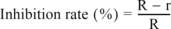
Where R is the radial growth of fungal mycelia on the control plate and r is the radial growth of fungal mycelia on the plate treated with AgNPs.
Results
Inhibition effect of AgNP
The inhibition effect of WA-CV-WA13B AgNPs at different concentrations was analyzed in PDA, MEA, and CMA (Table 3). In most cases, higher inhibition of fungal growth was recorded at a concentration of 100 ppm. In addition, most fungi showed growth inhibition with the increment of incubation time, and the inhibition showed similar patterns for each type of media. Absolute inhibition (100%) was observed on PDA medium against A-3, C-10, F-5, P-8, and P-9, and treatment with a 100 ppm concentration resulted in greater than 90% inhibition on PDA against C-1, D-1, G-1, M-1, and S-3. The lowest level of inhibition was observed against G-1 on PDA treated with a 10 ppm concentration of AgNPs (Table 3). On MEA, the highest level of inhibition was observed against M-1 (Table 3, Fig. 1) treated with a 25 ppm concentration of AgNPs (94.1%), and no inhibition was observed with treatment at 10 and 25 ppm concentrations of AgNPs against P-9. In addition, treatment with a 100 ppm concentration of AgNPs resulted in 90% inhibition against M-1 on MEA. Absolute inhibition (100%) was also observed on CMA treated with a 50 ppm concentration of AgNPs against C-10. The inhibition effect on CMA was greater than 90% against M-1 treated with a 100 ppm concentration of AgNPs and A-2 treated with 25 and 50 ppm concentrations of of silver nanoparticles, respectively (Table 3, Fig. 1). The lowest level of inhibition was observed against M-1 treated with a 10 ppm concentration of AgNPs on CMA (Table 3, Fig. 1). Therefore, the results suggested that maximum inhibition was obtained on PDA treated with a 100 ppm concentration of silver nanoparticles. Data on inhibition effects of WA-AT-WA13B AgNPs against various plant pathogens on PDA, MEA, and CMA are shown in Table 4. On PDA, absolute inhibition (100%) was obtained against A-3, F-4, M-1, and P-9 treated with a 100 ppm concentration of silver nanoparticles, and P-9 treated with a 50 ppm concentration of AgNPs (Table 4, Fig. 1). The lowest level of inhibition was observed against F-2 treated with 10 and 25 ppm concentrations of AgNPs on PDA, respectively. Likewise, on MEA, 100% inhibition was obtained against C-10 and P-8 treated with 50 and 100 ppm concentrations and P-9 with 25, 50, and 100 ppm concentrations of AgNPs; the lowest level of inhibition was observed against P-8 treated with a 10 ppm AgNPs solution (0.9%). In the case of CMA, 100% inhibition was observed against A-2, F-4, and F-5 treated with a 100 ppm concentration, as well as F-3 and F-4 treated with a 50 ppm concentration of silver nanoparticles. The lowest level of inhibition was observed against P-8 treated with a 10 ppm concentration of AgNPs on CMA (Table 4). Therefore, the results showed that in most cases, the inhibition effect increases with the increment of AgNP concentration (Table 4). The inhibition rate is also dependent on the culture media selected for the study.
Table 3.
Inhibitory rate (%) of silver nanoparticles WA-CV-WA13B against various plants pathogenic fungi on different media in vitro

Means followed by a different letter(s) in the same column differ significantly (p = 0.05) according to Duncan's multiple range test (DMRT).
PDA, potato dextrose agar; MEA, malt extract agar; CMA, corn meal agar.
aInhibition rates were determined based on five replicates of each experiment, inhibition rate of control = 0%.
Fig. 1.

Inhibition effect of silver nanoparticles (AgNPs) against Monosporascus cannonballus on PDA, MEA, and CMA in vitro. A, WA-CV-WA13B; B, WA-AT-WB13R; C, WA-PR-WB13R. PDA, potato dextrose agar; MEA, malt extract agar; CMA, corn meal agar; CTR, control.
Table 4.
Inhibitory rate (%) of silver nanoparticles WA-AT-WB13R against various plant pathogenic fungi on different media in vitro

Means followed by a different letter(s) in the same column differ significantly (p = 0.05) according to Duncan's multiple range test (DMRT).
PDA, potato dextrose agar; MEA, malt extract agar; CMA, corn meal agar.
aInhibition rates were determined based on five replicates of each experiment, inhibition rate of control = 0%.
Table 5 shows the inhibition effect of WA-PR-WB13R AgNPs against fungi on PDA, MEA, and CMA. On PDA, 100% inhibition was achieved against B-1, C-1, C-9, C-10, F-1, F-3, F-5, G-1, M-1, P-8, and P-9 treated with a 100 ppm concentration of AgNPs (Table 5, Fig. 1). In addition, greater than 90% inhibition was observed against other fungi treated with a 100 ppm concentration of AgNPs on PDA. The lowest level of inhibition was observed against P-8 treated with a 10 ppm concentration of AgNPs on PDA (Table 5). On MEA, 100% inhibition was observed against P-9 treated with 25, 50, and 100 ppm concentrations of AgNPs and C-10 cultured with a 25 ppm concentration of AgNPs (Table 5). The lowest level of inhibition was observed against F-3 treated with a 10 ppm concentration of AgNPs on MEA (20%). Similarly, a 100% inhibition effect of AgNPs was observed on CMA against P-9 treated with a 25 ppm concentration of AgNPs (Table 5). The lowest level of inhibition was observed against P-8 treated with a 10 ppm concentration of AgNPs. Therefore, the results suggested that inhibition rate was dependent on the concentration of AgNPs and culture media used in the study.
Table 5.
Inhibitory rate (%) of silver nanoparticles WA-PR-WB13R against various plant pathogenic fungi on different media in vitro

Means followed by a different letter(s) in the same column differ significantly (p = 0.05) according to Duncan's multiple range test (DMRT).
PDA, potato dextrose agar; MEA, malt extract agar; CMA, corn meal agar.
aInhibition rates were determined based on five replicates of each experiment, inhibition rate of control = 0%.
Discussion
Management of fungal diseases of food crops and fruits is economically important. Recently, a greater effort has been given to development of safe management methods that pose less danger to humans and animals, with a focus on overcoming deficiencies of synthetic fungicides. Findings from the current investigation demonstrated that AgNPs with low toxicity and a broad spectrum of antimicrobial activity were also very effective against plant phytopathogenic fungi. However, the current study is based on in vitro petri dish evaluation; therefore, extrapolation of these findings to more general cases is limited. Still, data from this study provide valuable preliminary efficacy data on silver compounds for use in control of plant pathogens. In this study, we analyzed the inhibition effect of three different AgNPs (WA-CV-WA13B, WA-AT-WB13R, and WA-PR-WB13R) against various plant pathogenic fungi in vitro. The results suggest that AgNPs are capable of inhibiting these pathogens; however, results vary according to the concentration and type of AgNPs applied to pathogens. Most fungi showed a high inhibition effect at a 100 ppm concentration of silver nanoparticles. In addition, results indicate that a higher inhibition rate was observed on PDA media, compared with others. Among AgNPs, WA-CV-WA13B showed the highest inhibition effect. In most cases, inhibition increased as the concentration of AgNPs increased. This could be due to the high density at which the solution was able to saturate and cohere to fungal hyphe and to deactivate plant pathogenic fungi. Reports on the mechanism of inhibitory action of silver ions on microorganisms have shown that upon treatment with Ag+, DNA loses its ability to replicate [10], resulting in inactivated expression of ribosomal subunit proteins, as well as certain other cellular proteins and enzymes essential to ATP production [11]. It has also been hypothesized that Ag+ primarily affects the function of membrane-bound enzymes, such as those in the respiratory chain [12, 13]. In summary, AgNPs exerted potent antifungal effects on fungi tested in vitro, probably through destruction of membrane integrity; therefore, it was concluded that AgNPs have considerable antifungal activity. Further investigation for field applications is needed.
Acknowledgements
This research was supported by a grant from the Ministry for Food, Agriculture, Forestry and Fisheries, and, in part, by the Agriculture and Life Sciences Research Institute (ALSRI) of Kangwon National University.
References
- 1.Jo YK, Kim BH, Jung G. Antifungal activity of silver ions and nanoparticles on phytopathogenic fungi. Plant Dis. 2009;93:1037–1043. doi: 10.1094/PDIS-93-10-1037. [DOI] [PubMed] [Google Scholar]
- 2.Clement JL, Jarrett PS. Antibacterial silver. Met Based Drugs. 1994;1:467–482. doi: 10.1155/MBD.1994.467. [DOI] [PMC free article] [PubMed] [Google Scholar]
- 3.Park HJ, Kim SH, Kim HJ, Choi SH. A new composition of nanosized silica-silver for control of various plant diseases. Plant Pathol J. 2006;22:295–302. [Google Scholar]
- 4.Chambers VW, Proctor CM, Kabler PW. Bactericidal effects of low concentrations of silver. J Am Water Works Assoc. 1962;54:208–216. [Google Scholar]
- 5.Kim JH. Nano silver chemotherapeutic agents and its applications. News Inf Chem Eng. 2004;22:655–660. [Google Scholar]
- 6.Berger TJ, Spadaro JA, Chapin SE, Becker RO. Electrically generated silver ions: quantitative effects on bacterial and mammalian cells. Antimicrob Agents Chemother. 1976;9:357–358. doi: 10.1128/aac.9.2.357. [DOI] [PMC free article] [PubMed] [Google Scholar]
- 7.Davies RL, Etris SF. The development and functions of silver in water purification and disease control. Catal Today. 1997;36:107–114. [Google Scholar]
- 8.Kim SW, Kim KS, Lamsal K, Kim YJ, Kim SB, Jung M, Sim SJ, Kim HS, Chang SJ, Kim JK, et al. An in vitro study of the antifungal effect of silver nanoparticles on oak wilt pathogen Raffaelea sp. J Microbiol Biotechnol. 2009;19:760–764. [PubMed] [Google Scholar]
- 9.Min JS, Kim KS, Kim SW, Jung JH, Lamsal K, Kim SB, Jung M, Lee YS. Effects of colloidal silver nanoparticles on sclerotium-forming phytopathogenic fungi. Plant Pathol J. 2009;25:376–380. [Google Scholar]
- 10.Feng QL, Wu J, Chen GQ, Cui FZ, Kim TN, Kim JO. A mechanistic study of the antibacterial effect of silver ions on Escherichia coli and Staphylococcus aureus. J Biomed Mater Res. 2000;52:662–668. doi: 10.1002/1097-4636(20001215)52:4<662::aid-jbm10>3.0.co;2-3. [DOI] [PubMed] [Google Scholar]
- 11.Yamanaka M, Hara K, Kudo J. Bactericidal actions of a silver ion solution on Escherichia coli, studied by energy-filtering transmission electron microscopy and proteomic analysis. Appl Environ Microbiol. 2005;71:7589–7593. doi: 10.1128/AEM.71.11.7589-7593.2005. [DOI] [PMC free article] [PubMed] [Google Scholar]
- 12.Bragg PD, Rainnie DJ. The effect of silver ions on the respiratory chains of Escherichia coli. Can J Microbiol. 1974;20:883–889. doi: 10.1139/m74-135. [DOI] [PubMed] [Google Scholar]
- 13.McDonnell G, Russell AD. Antiseptics and disinfectants: activity, action, and resistance. Clin Microbiol Rev. 1999;12:147–179. doi: 10.1128/cmr.12.1.147. [DOI] [PMC free article] [PubMed] [Google Scholar]