Figure 3.

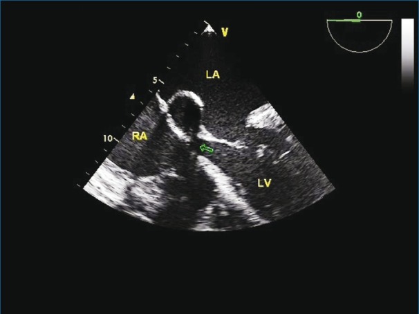
Transesophageal echocardiography in a four-chamber view showing aneurismal sac with neck (arrow) communicating with the LVOT

Transesophageal echocardiography in a four-chamber view showing aneurismal sac with neck (arrow) communicating with the LVOT