Figure 3.

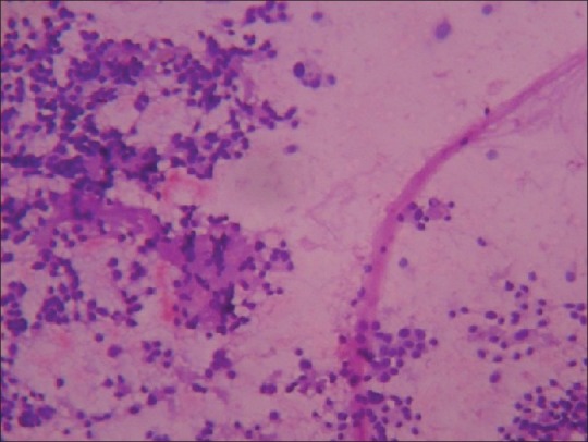
Photomicrograph of smear of medullary carcinoma thyroid, showing round cells with acellular eosinophilic material, (amyloid) in the background (Hematoxylin and Eosin stain, ×100)

Photomicrograph of smear of medullary carcinoma thyroid, showing round cells with acellular eosinophilic material, (amyloid) in the background (Hematoxylin and Eosin stain, ×100)