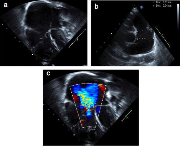
Figure 1.
Echocardiographic abnormalities in Menkes disease. A series of echocardiographic images from patient #3 at age two years who underwent balloon valvuloplasty in the newborn period for severe pulmonary valve stenosis. By age two years, the right ventricle, atrium (panel A), and pulmonary artery (panel B) were massively dilated, and there was severe tricuspid valve regurgitation (panel C). Although valvular insufficiency is not uncommon following valvuloplasty, the degree of right heart dilation and dysfunction in this patient with Menkes disease was more severe, and developed more rapidly than expected.

