Figure 2.
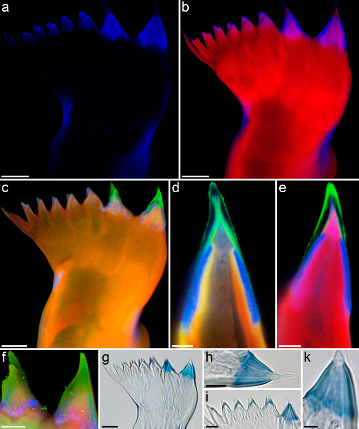
(a–f) Confocal laser scanning micrographs of mandibular gnathobases from female Centropages hamatus, cranial view ([a–c] maximum intensity projections [MIPs] showing the whole gnathobase; [d, e] 1 µm thick optical sections through the ventral tooth; [f] MIP showing the ventral and the first central tooth): (a) distribution of resilin; (b) chitinous exoskeleton (red) and resilin-dominated structures (blue); (c–f) chitinous exoskeleton (orange, red), resilin-dominated structures (blue, light blue) and silica-containing structures (green). (g–k) Bright-field micrographs of mandibular gnathobases from female C. hamatus stained with toluidine blue, cranial view: (g) overview of a whole gnathobase; [h, k] detailed view of the ventral tooth; (i) detailed view of the first central tooth and the smaller teeth in the central and dorsal parts of the gnathobase. Scale bars = 20 µm (a, b, c, g), 10 µm (f, h, i), 5 µm (d, e, k).

