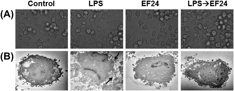
Fig. 4.
Morphological changes induced by LPS treatment on JAWS II DCs are abrogated by EF24. (A) Light microscopic pictures of DCs treated with vehicle-control, 100 ng ml−1 LPS, 10 μM EF24 and 100 ng ml−1 LPS followed by 10 μM EF24 (LPS→EF24). The pictures were acquired after 5 h of treatment. (B) Transmission electron photomicrographs (×3000) of JAWS II DCs treated similarly. The arrows indicate the conspicuous dendrites on the mature JAWS II DCs that are subdued by EF24 treatment. The electron micrographs are representative of two independent experiments.

