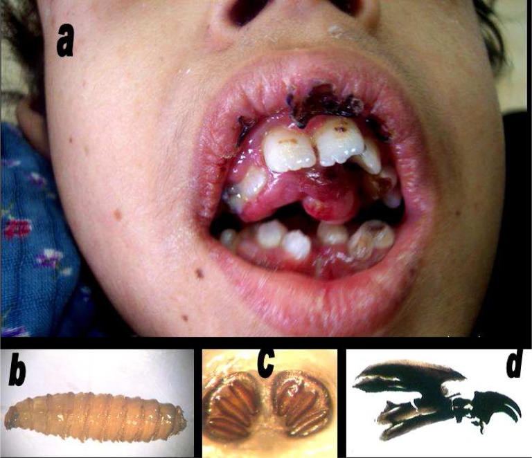
Fig. 1.
(a): The patient’s mouth part infested by Chrysomya bezziana larva, (b): C. bezziana in the third stage of larvae (125×), (c): Posterior spiracle of C. bezziana in the third stage of larvae (125×), (d): Cephalopharyngeal skeleton of C. bezziana in the third larval stage (125×) (Original photos)

