
Figure 4.
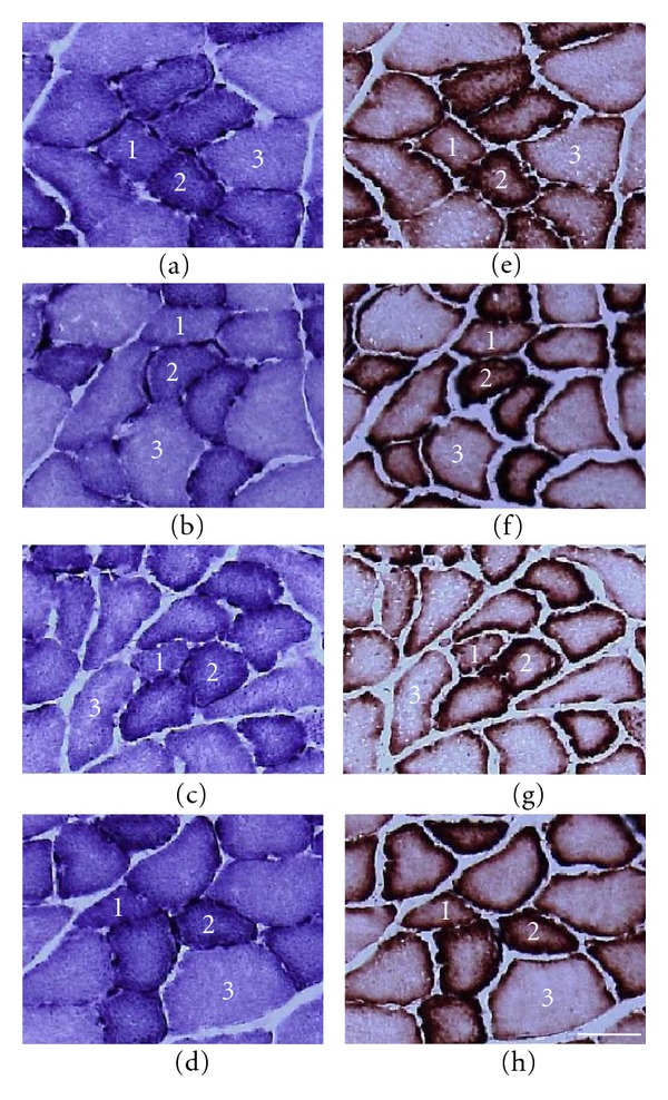
Transverse sections of the extensor digitorum longus muscle assayed for SDH ((a)–(d)) and cytochrome c ((e)–(h)) staining. (a) and (e) LC group; (b) and (f) LH group; (c) and (g) OC group; (d) and (h) OH group. 1: type I fiber; 2: type IIA fiber; 3: type IIB fiber. Bar = 50 μm.