
Fig. 2.
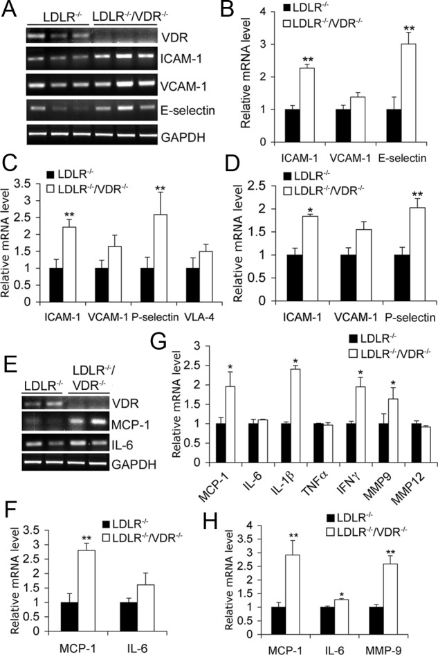
Expression of adhesion molecules and inflammatory cytokines in aorta and macrophages. A and B, RT-PCR amplification (A) and quantitation (B) of adhesion molecule transcripts in the aorta isolated from LDLR−/− and LDLR−/−/VDR−/− mice fed the HFHC diet for 4 wk. C and D, Real-time RT-PCR quantitation of adhesion molecules in LDLR−/− and LDLR−/−/VDR−/− peritoneal macrophages at baseline (C) or stimulated with 50 μg/ml acLDL for 24 h (D). E and F, RT-PCR amplification (E) and quantitation (F) of MCP-1 and IL-6 levels in aorta isolated from 4-wk HFHC diet-treated LDLR−/− and LDLR−/−/VDR−/− mice. G and H, Real-time RT-PCR quantitation of cytokines and chemokines in LDLR−/− and LDLR−/−/VDR−/− peritoneal macrophages at baseline (G) or after being stimulated with acLDL (H). *, P < 0.05 vs. LDLR−/−; n = 5–10. GAPDH, Glyceraldehyde-3-phosphate dehydrogenase; IFN, interferon; MMP, matrix metalloproteinase; VCAM, vascular cell adhesion molecule.