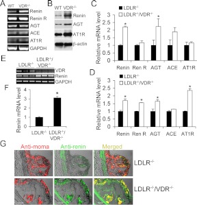
Fig. 5.
Effects of VDR deletion on the local RAS in macrophages and aorta. A and B, Assessment of RAS components in peritoneal macrophages isolated from WT and VDR(−/−) mice by RT-PCR (A) and Western blotting (B). C and D, Real-time RT-PCR quantitation of RAS components in peritoneal macrophages isolated from LDLR−/− and LDLR−/−/VDR−/− mice at baseline (C) or after acLDL stimulation (D). E and F, RT-PCR amplification (E) and quantitation (F) of renin transcripts in the aorta from LDLR−/− and LDLR−/−/VDR−/− mice. G, Atherosclerotic lesions from LDLR−/− and LDLR−/−/VDR−/− mice stained with anti-MOMA-2 (red) and anti-renin (green) antibodies. Note that renin is colocalized with macrophages (yellow), and LDLR−/−/VDR−/− macrophages have higher renin levels compared with LDLR−/− macrophages. ACE, Angiotensin-converting enzyme; GAPDH, glyceraldehyde-3-phosphate dehydrogenase; Re R, renin receptor.

