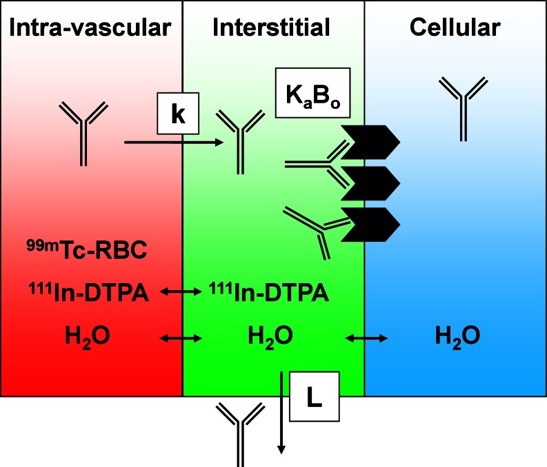
Fig. 3.
Adapted with permission from Boswell et al. (36). Tissue compartments and the radioactive tracers used to assess their physiological parameters. Each tissue can be separated into three compartments: intravascular, interstitial, and cellular. Technetium-99m-labeled red blood cells (99m Tc-RBC) and 111In-DTPA allow measurement of vascular and extracellular volumes, respectively. The antibody’s receptor, if present, may be expressed on the cell surface, exposed to the interstitial fluid. An antibody in circulation may extravasate from blood into interstitial space at a rate (k), where it may encounter a number (B o) of receptors for which it has binding affinity (K a). The antibody may also return to circulation via lymphatic flow (L)

