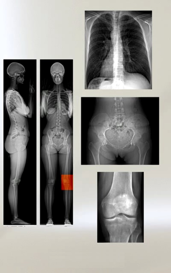
Fig. 2.
Two-dimensional digital X-ray images captured by the EOS™ 2D/3D system. From left to right and top to bottom: full-body biplanar lateral and posterioanterior (PA) images; PA thorax; PA pelvis; and PA right knee images. 1:1 scale, high contrast, high dynamic range, high resolution images free of distortion or artifacts. (Illustration used with permission from EOS Imaging, Paris, France)

