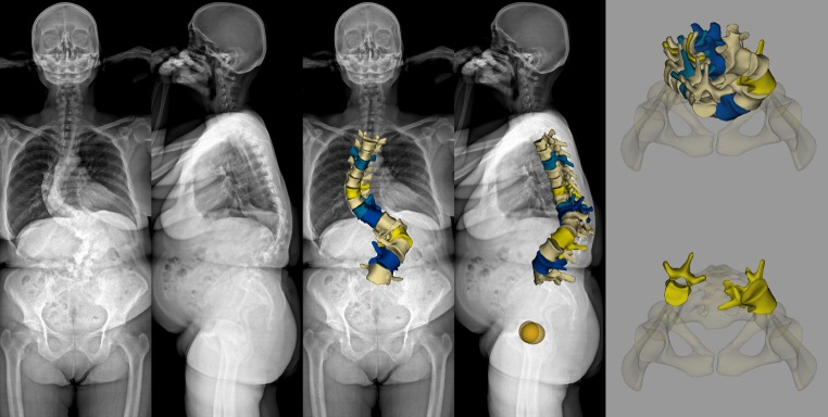
Fig. 6.
EOS™ 2D examination and sterEOS 3D reconstruction of the spine in adult degenerative scoliosis. A 59-year-old female patient with a history of degenerative lumbar spine deformity diagnosed five years earlier, causing low back pain and sciatica in her right upper leg. Anterioposterior and lateral EOS 2D images (image 1-2 from left) show a degenerative scoliosis consisting of a large left convex thoracolumbar curve with a significant apical vertebral rotation and a right convex thoracic curve. The upper body seems to be compensated and the pelvis position appears normal. EOS™ 3D reconstructed spine model of spine region Th4-L5 overlayed on the X-ray images (image 3-4 from left) demonstrates the 3D depiction of the deformity in frontal and sagittal plane. End-vertebrae Th5, Th11(azure) and apical vertebra Th8 (yellow) of the 90° thoracic curve and end-vertebrae Th12, L4 (blue) and apical vertebra L2 (yellow) of the 110° thoracolumbar curve are highlighted in colour. Sagittal curves appear abnormal with an apparent angular kyphosis of the lumbar region (L1-L5 lordosis -4°). Relative lateral deviations and rotational changes of vertebrae are more apparent in the horizontal plane top view image showing the reconstructed 3D spine model with the pelvis (upper right). Axial rotation of apical vertebrae L2 and Th8 (53° and −20°, respectively) with their lateral translation and relative position are more conclusive when visualised individually with the pelvis (bottom right)

