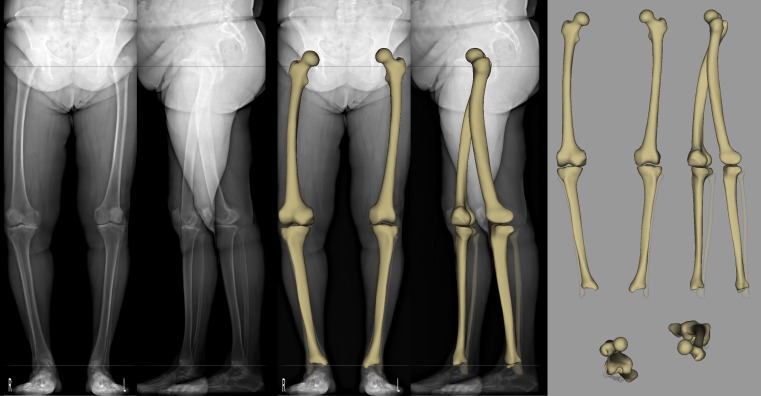
Fig. 7.
EOS™ 2D examination and sterEOS 3D reconstruction of the lower limbs in adult degenerative knee arthritis. A 72-year-old female patient with a history of right knee problems showing an increasing pain in the last two years. Physical examination revealed antalgic gait with a limp, 10° flexion contracture, 15° varus position, knee flexion reduced to 110° and subpatellar crepitation. Biplanar EOS™ X-ray images (image 1-2 from left) demonstrate spondylosis and spondylarthritis in visible parts of the lumbar spine, intermediate arthritic aberrations of the hips and both knees with emphasis in the medial compartment and lateralisation of both patella. Apexes of patellae are spiculated and osteophytes are visible in both femoropatellar surfaces. Varus position of the right knee is more apparent. EOS™ 3D reconstructed 3D models of the lower limbs overlayed to the X-ray images (image 3-4 from left) and shown in frontal, sagittal and horizontal plane top views (image 5-6-7 on the right) resulted in the following relevant clinical parameters: knee varus – right: 13°, left: 6°; femoral mechanical angle – right: 87°, left: 92°; tibial mechanical angle – right: 82°, left: 87°; HKS angle – right: 4.0°, left: 6.0°

