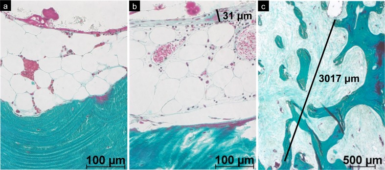
Fig. 7.
Bone–cement interface in retrieved hip-resurfacing arthroplasty (HRA) specimens: a Specimen with viable bone and absent fibrous interface membrane. A discrete layer of macrophages was visible on the surface of the cement (plastic embedding; stain: Goldner trichrome, original magnification ×200). b Thin fibrous membrane on the bone–cement interface (plastic embedding; stain: Goldner trichrome, original magnification ×200). c Thick fibrous membrane (mid) at the border of advanced osteonecrosis (above; plastic embedding; stain Goldner trichrome, original magnification ×25)

