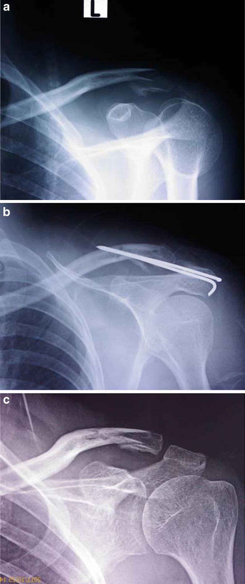
Fig. 2.
a X-ray anteroposterior view (left shoulder) showing displaced type 2 lateral end clavicle fracture. b Immediate post operative X-ray showing reduced fracture with transacromial Kirshner wire. The drill hole for figure-eight suture is well marked in both the fragments. c Well reduced and united fracture lateral end clavicle after Kirshner wire removal at six-months follow-up

