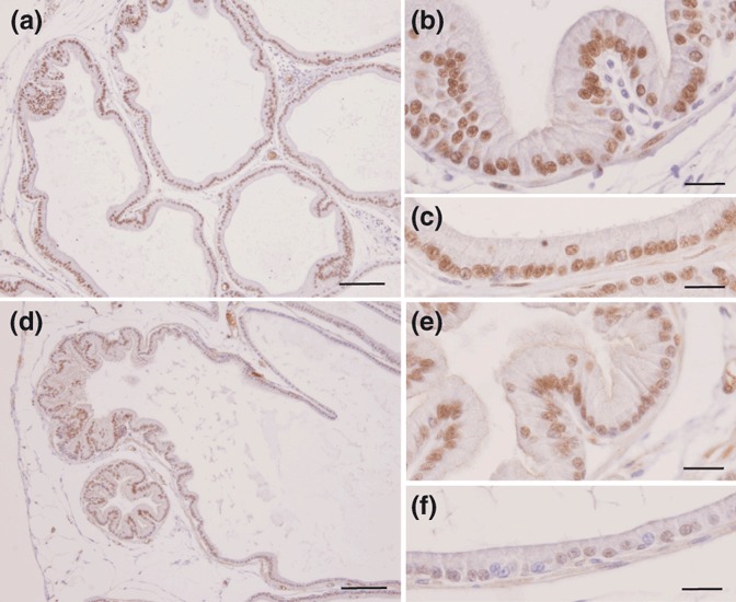
Figure 6.

Immunocytochemistry for AR in the ventral prostate of control rats (a–c) or rats treated with DEX (d–f). Distal region (a, b, d and e) and intermediate region of the acini (c and f). Note the reduction in the staining of the AR in the prostate epithelium of the DEX-treated group and the decreasing intensity of nuclear staining for this receptor after treatment with DEX. Scale bars = 100 μm for a and d; 20 μm for b, c, e and f.
