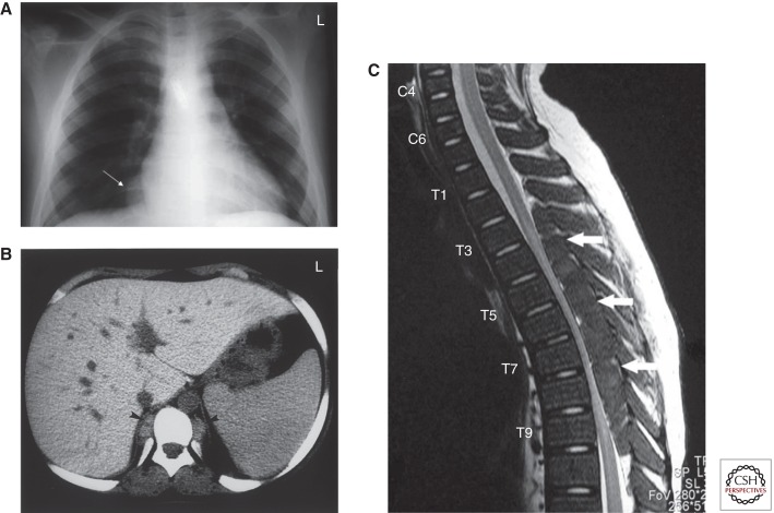
Figure 3.
Paraspinal extramedullary hematopoietic pseudotumors. (A) Chest X-ray showing expanded anterior rib ends consistent with medullary hyperplasia. A paraspinal mass is seen in the right lower zone (white arrow). (B) Computed tomography scan showing inactive paraspinal extramedullary hematopoietic lesion with increased density compared with soft tissue, caused by iron deposition (black arrowheads). (C) Magnetic resonance image of cervical and thoracic spine. T2-weighted sagittal image showing thoracic cord compression by extramedullary intraspinal epidural hematopoietic mass from T2 to T10 (white arrows). (From Haidar et al. 2010; reprinted, with permission.)

