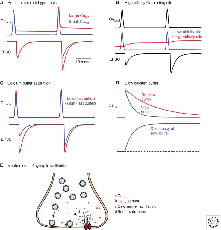
Figure 3.
Proposed mechanisms of facilitation. (A) The residual calcium hypothesis based on a single type of low-affinity calcium sensor is shown for two types of calcium signals. When the residual calcium signal is a significant fraction of the local calcium signal, significant facilitation occurs (red traces), but when the residual calcium signal is much smaller than the calcium signal near the calcium channel, this mechanism results in very little enhancement (blue traces). (B) Another type of residual calcium model is shown that is based on two types of calcium sensors, a fast, low-affinity sensor and a slow, high-affinity sensor. The residual calcium signal can activate the high-affinity receptor to produce facilitation. (C) A comparison of the calcium signals and resulting EPSCs for a synapse in which a presynaptic bouton contains either a high concentration (blue) or a low concentration (red) of a rapid calcium buffer illustrates another mechanism of facilitation. (D) A slow calcium buffer binds presynaptic calcium slowly and by doing so accelerates the decay of presynaptic calcium, which can in turn affect facilitation. (E) Schematic illustrating mechanisms of facilitation.

