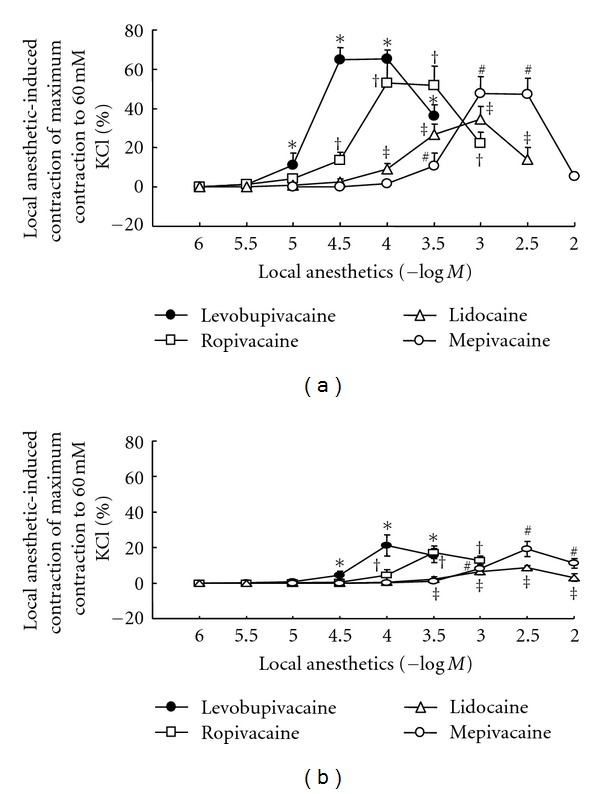
Figure 1.

Concentration-response curves induced by levobupivacaine, ropivacaine, lidocaine, and mepivacaine in isolated endothelium-denuded (a) and -intact (b) aorta. All values are shown as mean ± SD and expressed as the percentage of the maximal contraction induced by 60 mM KCl. N indicates the number of rats from which descending thoracic aortic rings were derived. (a) Isotonic 60 mM KCl-induced contraction in endothelium-denuded aorta: 100% = 2.94 ± 0.66 g (n = 6) with levobupivacaine, 100% = 3.24 ± 0.51 g (n = 6) with ropivacaine, 100% = 2.88 ± 0.49 g (n = 6) with lidocaine, and 100% = 2.91 ± 0.36 g (n = 6) with mepivacaine. *P < 0.01 versus 10−6 M levobupivacaine; †P < 0.01 versus 10−6 M ropivacaine; ‡P < 0.01 versus 10−6 M lidocaine; #P < 0.01 versus 10−5 M mepivacaine. (b) Isotonic 60 mM KCl-induced contraction in endothelium-intact aorta: 100% = 2.42 ± 0.50 g (n = 6) with levobupivacaine, 100% = 2.44 ± 0.43 g (n = 6) with ropivacaine, 100% = 2.11 ± 0.56 g (n = 6) with lidocaine, and 100% = 2.43 ± 0.28 g (n = 6) with mepivacaine. *P < 0.001 versus 10−6 M levobupivacaine; †P < 0.01 versus 10−6 M ropivacaine; ‡P < 0.05 versus 10−6 M lidocaine; #P < 0.001 versus 10−5 M mepivacaine.
