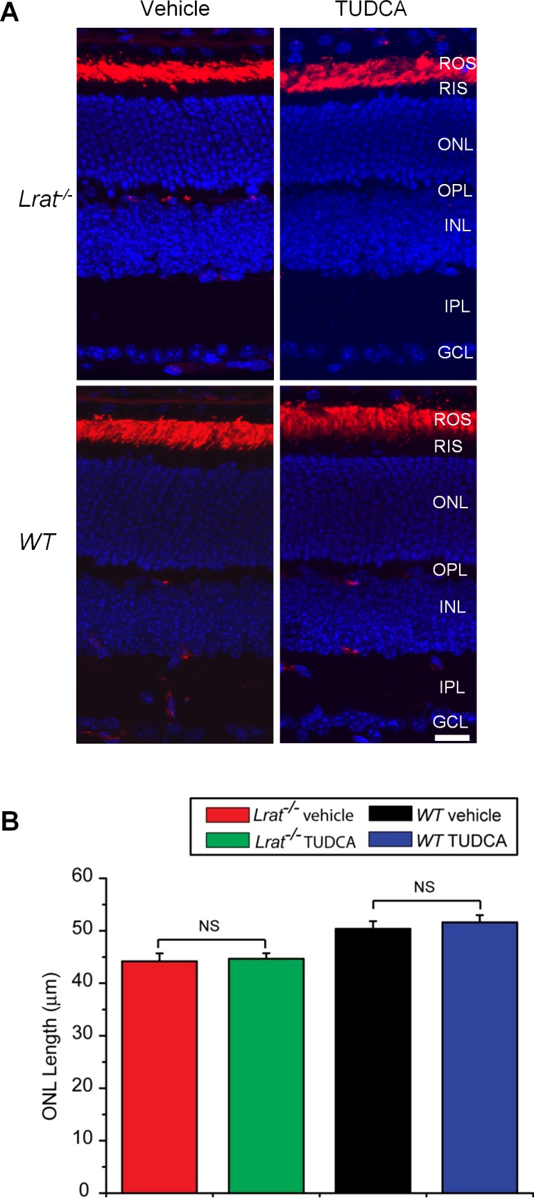
Figure 3. .

Effect of TUDCA treatment on rod photoreceptors. (A) P28 Lrat–/– and WT retinal sections were labeled with rhodopsin antibody 1D4 (in red). Nuclei were stained with DAPI (blue). Scale bar = 20 μm. (B) ONL length was measured from TUDCA-injected Lrat–/– (n = 8), vehicle-injected Lrat–/– (n = 6), TUDCA-injected WT (n = 6), and vehicle-injected WT (n = 6). Data were presented as mean ± SEM. NS, not significant.
